Breast Anatomy Quadrants - Healthy Breast With Ultrasound Radiology Key. Understanding normal breast anatomy and its lymphatic drainage can also help us evaluate the extent of cancers more accurately. A wide variation of homogeneously dense, milky structures (representing glandular tissue) interrupted by areas of curved or round radiolucent fat Whether tumour location is prognostic is unclear. Rotate into a sagittal plane and repeat the pattern. Lateral quadrants of the breast;
The muscles involved include the pectoralis major, serratus anterior, external oblique, and rectus abdominis fascia. Rotate into a sagittal plane and repeat the pattern. The breast is located on the anterior thoracic wall in the superficial fascia of the pectoral region. Behind, beneath, under, underneath, next to, above, cephalad to, or below nipple. C502 upper inner quadrant (uiq) of breast.
Anatomy Of The Breast Medcaretips Com from medcaretips.com The glands function is provided by hormones estrogen. Polythelia (supranumerary nipple) polymastia (accessory breast tissue) radiographic appearance mammography. C502 upper inner quadrant (uiq) of breast. Mainly to the axillary lymph nodes as mentioned above. Both malignant and benign breast disease may arise in these ectopic sites. Paget disease with underlying tumor. C501 central portion of breast (subareolar) area extending 1 cm around areolar complex. These lobules are separated by fibrous septa running from the subcutaneous tissues to the fascia of the chest wall.
Repeat this across the breast.
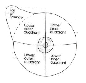
C501 central portion of breast (subareolar) area extending 1 cm around areolar complex. Tumour location within the breast varies with the highest frequency in the upper outer quadrant (uoq) and lowest frequency in the lower inner quadrant (liq). They rest on the major chest muscle, the pectoralis major. Anatomy of the breast situation. Behind, beneath, under, underneath, next to, above, cephalad to, or below nipple. Understanding normal breast anatomy and its lymphatic drainage can also help us evaluate the extent of cancers more accurately. Whether tumour location is prognostic is unclear. The breast is divided into four quadrants: Anatomy of the breast 1. The muscles involved include the pectoralis major, serratus anterior, external oblique, and rectus abdominis fascia. They are localized in the front of the breast within the 3 to 7 rib. Both malignant and benign breast disease may arise in these ectopic sites. It often extends to the axillary tail (tail.
This illustration shows the makeup of breast anatomy both inside and outside. Repeat this across the breast. Code laterality to the side with the positive nodes. Each lobe is comprised of many lobules, at the end of which are tiny bulblike glands, or sacs, where milk is produced in response to hormonal signals. C502 upper inner quadrant (uiq) of breast.
Anatomy Of The Breast Medcaretips Com from medcaretips.com C501 central portion of breast (subareolar) area extending 1 cm around areolar complex. Each breast contains 15 to 20 lobes arranged in a circular fashion. Lateral quadrants of the breast; The breast is divided into four quadrants: 2:00 in the right breast is in the uiq, whereas 2:00 in the left breast is in the uoq. They are present in both males and females, yet are more prominent in females following puberty. Anatomy of the breast yapa wijeratne faculty of medicine university of peradeniya 2. Glandular tissue is most abundant in upper outer quadrant of breast;
This illustration shows the makeup of breast anatomy both inside and outside.
Code the primary site to c509 when there are multiple tumors (two or more) in at least two quadrants of the breast. C502 upper inner quadrant (uiq) of breast. Paget disease with underlying tumor. Anatomy of the breast situation. They are present in both males and females, yet are more prominent in females following puberty. The breast is a mound of fibrous stroma with adipose, ductal, and glandular tissue overlying the anterior chest wall ( fig. These lobules are separated by fibrous septa running from the subcutaneous tissues to the fascia of the chest wall. Rotate into a sagittal plane and repeat the pattern. Each lobe is comprised of many lobules, at the end of which are tiny bulblike glands, or sacs, where milk is produced in response to hormonal signals. A variation, particularly in larger or mobile breasts, is to apply the grid pattern quadrant by quadrant. The breast is divided into four quadrants: C501 central portion of breast (subareolar) area extending 1 cm around areolar complex. Anatomy of the breast normal anatomy.
Each breast contains 15 to 20 lobes arranged in a circular fashion. As a result, half of all breast cancers occur here. 2:00 in the right breast is in the uiq, whereas 2:00 in the left breast is in the uoq. Whether tumour location is prognostic is unclear. Anatomy of the breast normal anatomy.
Pdf Efficacy Of Combined Mammographic And Sonographic Evaluation Of Both Dense And Fatty Breast from www.researchgate.net C50.6 is the code for axillary tail or tail of breast. The breast is a mound of fibrous stroma with adipose, ductal, and glandular tissue overlying the anterior chest wall ( fig. C502 upper inner quadrant (uiq) of breast. The breast is located on the anterior thoracic wall in the superficial fascia of the pectoral region. Anatomy of the breast yapa wijeratne faculty of medicine university of peradeniya 2. They are localized in the front of the breast within the 3 to 7 rib. As a result, half of all breast cancers occur here. Move across and repeat the sweep inferior to superior.
Rotate into a sagittal plane and repeat the pattern.
Laterality laterality must be coded for all subsites. « previous (anatomy) next (regional lymph nodes) ». A wide variation of homogeneously dense, milky structures (representing glandular tissue) interrupted by areas of curved or round radiolucent fat Code laterality to the side with the positive nodes. Fat accounts for its smooth contour and most of its bulk. C50.6 is the code for axillary tail or tail of breast. Tumour location within the breast varies with the highest frequency in the upper outer quadrant (uoq) and lowest frequency in the lower inner quadrant (liq). Glandular tissue is most abundant in upper outer quadrant of breast; The breast is a modified sweat gland located in the superficial fascia of the anterior chest wall.the major portion of the breast tissue is situated between the second and third rib superiorly, the sixth and seventh costal cartilage inferiorly, the anterior axillary line laterally, and the sternal border medially. Understanding normal breast anatomy and its lymphatic drainage can also help us evaluate the extent of cancers more accurately. Polythelia (supranumerary nipple) polymastia (accessory breast tissue) radiographic appearance mammography. A layer of fat surrounds the glands and extends throughout the breast. The breast lies over the musculature that encases the chest wall.
Rotate into a sagittal plane and repeat the pattern anatomy quadrants. The breast is divided into four quadrants:
Share :
Post a Comment
for "Breast Anatomy Quadrants - Healthy Breast With Ultrasound Radiology Key"


Post a Comment for "Breast Anatomy Quadrants - Healthy Breast With Ultrasound Radiology Key"